
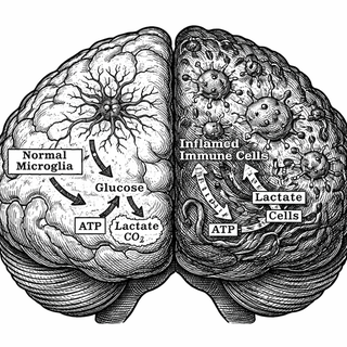
Deep within the folds of the human brain, microglia - the nervous system's resident security guards - are supposed to be keeping the peace. They patrol the tissue, tidying up debris, checking IDs at the blood-brain barrier, and generally making sure everything runs smoothly. But in multiple sclerosis, these once-loyal sentinels turn traitor. They flip a metabolic switch, start guzzling glucose like teenagers raiding the fridge at midnight, and begin churning out inflammatory molecules that strip the protective myelin coating from nerve fibers. The worst part? Until now, we've had a remarkably hard time catching them in the act.
A new clinical trial out of the University of Virginia (NCT07510607) is aiming to change that, and the technology they're using sounds like it was borrowed from a science fiction movie: hyperpolarized carbon-13 MRI.
What Exactly Is Hyperpolarized Carbon-13 MRI?
Standard MRI gives us beautiful pictures of brain structure. It can show us lesions, atrophy, and all the anatomical scars that MS leaves behind. But here's the frustrating thing - by the time those scars show up on a regular scan, the damage is already done. It's like detecting a fire by photographing the ashes.
Hyperpolarized carbon-13 MRI takes a fundamentally different approach. Instead of looking at structure, it watches metabolism in real time. Here's the gist: researchers take pyruvate (a molecule your cells use for energy), tag it with carbon-13, and then "hyperpolarize" it - essentially giving the carbon atoms a massive magnetic boost that makes them 10,000 to 100,000 times more visible on MRI. They inject this supercharged pyruvate into the bloodstream, and then they watch what the brain does with it.
In healthy tissue, cells politely process pyruvate through oxidative phosphorylation - the slow, efficient, well-mannered energy pathway. But inflamed immune cells? They've switched to glycolysis, a faster and messier metabolic strategy that rapidly converts pyruvate to lactate. This metabolic rebellion is called the Warburg effect, and it makes activated microglia and infiltrating macrophages light up like a neon sign on the scan.
The Warburg Effect: Not Just for Cancer Anymore
If the Warburg effect sounds familiar, you might be thinking of cancer biology - and you'd be right. Otto Warburg first described this metabolic quirk in tumor cells back in the 1920s, and it's been the basis for PET scans ever since. What's become increasingly clear over the past decade is that activated immune cells pull the exact same trick. When microglia and macrophages shift into attack mode, they reprogram their metabolism toward aerobic glycolysis, prioritizing speed over efficiency to fuel their inflammatory rampage (O'Neill et al., Nature Reviews Immunology, 2016; DOI: 10.1038/nri.2016.70).
Preclinical research has already demonstrated that hyperpolarized carbon-13 imaging can detect this metabolic shift in animal models of MS. Guglielmetti and colleagues showed that increased hyperpolarized lactate production in the brain correlated directly with microglial and macrophage infiltration in a mouse model of neuroinflammation (Guglielmetti et al., PNAS, 2017; DOI: 10.1073/pnas.1613345114). The signal was specific, measurable, and - most excitingly - changed with treatment response.
While hyperpolarized carbon-13 MRI has been used in human oncology studies, this trial represents one of its very first applications to neuroinflammation in living, breathing humans. That alone makes this worth paying attention to.
What the Trial Actually Involves
The study focuses on patients with relapsing-remitting MS - the most common form of the disease, characterized by episodes of new or worsening symptoms followed by periods of partial or complete recovery. Participants will undergo hyperpolarized carbon-13 MRI scans alongside conventional MRI, clinical assessments, and blood biomarker analysis.
The key question: can this metabolic imaging predict who will respond to anti-CD20 disease-modifying therapy? Anti-CD20 agents like ocrelizumab and ofatumumab work by depleting B cells, and they've transformed MS treatment over the past several years (Hauser et al., New England Journal of Medicine, 2020; DOI: 10.1056/NEJMoa1917246). But here's the rub - not every patient responds equally, and we don't have a great way to predict who will benefit most. Imagine being able to look at a patient's brain metabolism before starting treatment and say, "Based on your inflammatory profile, this therapy has an excellent chance of working for you." That's the kind of precision medicine this trial is reaching toward.
Why This Matters for Patients
For the roughly 2.8 million people living with MS worldwide, disease monitoring is a perpetual frustration. Conventional MRI can show new lesions, but lesion counts don't always correlate with how patients actually feel or how their disease is progressing. There's a growing recognition that "smoldering" neuroinflammation - chronic, low-grade immune activation driven largely by innate immune cells - plays a major role in the progressive disability that accumulates over time (Absinta et al., Nature, 2021; DOI: 10.1038/s41586-021-03892-7).
This is exactly the kind of inflammation that hyperpolarized carbon-13 MRI could reveal. Standard scans might show a brain that looks stable, while metabolically, the microglia are quietly simmering away. Wouldn't it be something if we could detect that simmering before it boils over?
And the applications don't stop at diagnosis. If this imaging technique works as hoped, it could fundamentally change how we monitor treatment response. Instead of waiting months or years to see whether new lesions appear - essentially waiting for failure - clinicians could potentially track metabolic changes within weeks, adjusting therapy before irreversible damage occurs.
The Bigger Picture
Grist and colleagues have already demonstrated that hyperpolarized carbon-13 MRI can map brain metabolism in healthy humans, establishing the baseline measurements needed to detect disease-related changes (Grist et al., NeuroImage, 2019; DOI: 10.1016/j.neuroimage.2019.116085). Building on that foundation, this trial asks whether we can use the same technology to peer into the metabolic chaos of neuroinflammation.
If successful, the implications extend well beyond MS. Microglial activation and the associated metabolic shift play roles in Alzheimer's disease, Parkinson's disease, traumatic brain injury, and a growing list of neurological conditions. A non-invasive tool that can visualize innate immune activation in the living human brain? That's not just a better diagnostic - it's a new window into how the brain fights, fails, and sometimes turns against itself.
Will this single trial solve MS? Of course not. But it represents the kind of creative, boundary-pushing science that moves the needle. Sometimes the most important discoveries don't come from new drugs, but from new ways of seeing.
For more details on trial design, eligibility, and enrollment, visit the ClinicalTrials.gov study page or review the study details table.
Disclaimer: This blog post is for informational and educational purposes only and does not constitute medical advice. Clinical trials are experimental by nature, and outcomes are not guaranteed. Always consult a qualified healthcare provider for medical decisions. Trial information sourced from ClinicalTrials.gov (NCT07510607).