
Let's be honest - when most people think about glamorous 3D imaging technology, they're picturing movie special effects or maybe some fancy architectural visualization. Nobody wakes up thinking, "You know what would be really cool? A 3D ultrasound of my armpit." And yet, here we are, in a timeline where researchers at Mayo Clinic are doing exactly that - and it might just save lives.
The clinical trial NCT05704283 is investigating 3D ultrasound imaging for axillary lymph nodes in patients with breast cancer. If you're wondering why anyone would care about lymph nodes in your underarm area, well, buckle up. Those little bean-shaped things might be the most important real estate in breast cancer staging, and figuring out what's happening there without surgery is kind of a big deal.
The Armpit Problem (Yes, That's a Technical Term Now)
Here's the situation: when breast cancer is diagnosed, one of the first questions oncologists need to answer is whether the cancer has spread to the lymph nodes in the armpit - technically called the axillary lymph nodes. These nodes act like little checkpoint stations for your immune system, which means they're often the first place cancer cells show up when they start traveling.
Knowing whether cancer has reached these nodes changes everything about treatment planning. If they're clear, that's a much better prognosis. If they're involved, the treatment strategy needs to be more aggressive. Traditionally, the only way to know for sure was to surgically remove some nodes and examine them under a microscope. This procedure, called sentinel lymph node biopsy, is pretty standard - but it's still surgery, which means anesthesia, incisions, recovery time, and the potential for complications like lymphedema (chronic arm swelling that approximately zero patients enjoy).
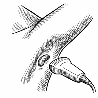
What if we could get that information without cutting anyone open? That's where 3D ultrasound enters the chat.
From Fuzzy Blobs to Actually Useful Images
Standard ultrasound has been used to look at lymph nodes for decades. The technology is cheap, widely available, doesn't involve radiation, and can be done right in the doctor's office. The problem? Traditional ultrasound images are... let's say "interpretive." They're 2D slices through a 3D structure, which means the person reading them needs to mentally reconstruct what they're looking at. It's like trying to understand the layout of a house by looking at a few random photographs taken through the windows.
3D ultrasound changes the game by capturing a volume of tissue and reconstructing it as an actual three-dimensional image. Instead of squinting at grainy slices and hoping you're interpreting them correctly, clinicians can rotate the image, zoom in on specific areas, and see the actual shape and structure of lymph nodes. It's the difference between reading a map and using Google Earth.
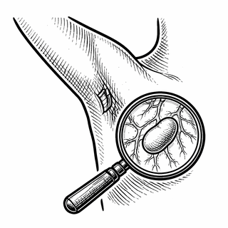
The Mayo Clinic trial is building on this technology by combining 3D ultrasound with other modalities - specifically B-mode imaging, microvessel imaging, and shear wave elastography. If that sounds like a lot of technical jargon, here's the translation: they're looking at the structure (B-mode), the blood vessel patterns (microvessel imaging), and how stiff the tissue is (elastography). Cancerous lymph nodes tend to have all sorts of weird characteristics - abnormal blood vessel patterns, different stiffness, altered shapes - and this approach captures multiple red flags simultaneously.
The AI Factor
Now here's where it gets really interesting. Mayo Clinic researchers have developed something called quantitative high-definition microvessel imaging (q-HDMI), which can capture images of blood vessels as small as 150 microns - about twice the width of a human hair. That's incredibly detailed, but also creates massive amounts of data. More data than any human could reasonably analyze in a clinical timeframe.
Enter machine learning. The researchers have identified specific biomarkers from these images - characteristics of tiny vessels like shape, pattern, irregularity, and complexity - and trained algorithms to sort the data into benign or malignant categories. According to Mayo's own reporting, this approach has achieved "nearly 100% accuracy" in determining malignant versus benign masses, regardless of tumor size.
I should note that "nearly 100%" in a research context usually comes with asterisks and caveats about small sample sizes, controlled conditions, and the need for further validation. But even if real-world performance is somewhat lower, we're talking about a non-invasive technique approaching surgical biopsy accuracy. That's remarkable.
Earlier studies on computer-aided diagnosis (CAD) systems for 3D volumetric ultrasound showed that radiologists using CAD improved their performance from an area under the ROC curve of 0.83 to 0.90 for distinguishing malignant from benign masses. For context, 1.0 would be perfect accuracy, and 0.5 would be random guessing. Moving from 0.83 to 0.90 represents a meaningful improvement in diagnostic confidence.
Why This Matters Beyond the Cool Factor
The practical implications of accurate non-invasive lymph node assessment are substantial. First, there's the obvious benefit of potentially avoiding unnecessary surgeries. If 3D ultrasound can reliably identify nodes that are definitely benign, those patients might be able to skip sentinel lymph node biopsy entirely. That's fewer surgical procedures, lower healthcare costs, and - most importantly for patients - less time spent dealing with surgical recovery instead of living their lives.
Second, there's the issue of surgical timing and planning. When surgery is necessary, knowing exactly which nodes are involved helps surgeons plan their approach. Going in with a detailed 3D map is considerably better than the surgical equivalent of "we'll see what we find."
Third, the technology could improve monitoring during treatment. For patients receiving neoadjuvant therapy (treatment given before surgery), being able to track lymph node changes non-invasively could help oncologists assess whether the treatment is working without requiring repeated biopsies.
The Bigger Picture in Breast Cancer Care
This trial fits into a broader movement toward less invasive breast cancer management. The NAUTILUS trial (NCT04303715), for example, is investigating whether sentinel lymph node biopsy can be safely omitted in certain clinically node-negative patients. The thinking is that if imaging can reliably rule out lymph node involvement, maybe we don't need to surgically confirm it in every case.
Ultrasound has been a workhorse in breast cancer detection and management for decades. It's simple, cheap, and widely available - unlike MRI, which requires specialized equipment and significant time. Research has shown that real-time MR-US navigation techniques can achieve detection rates of 90.7% for breast lesions and axillary lymph nodes, compared to 43% for conventional ultrasound alone. The diagnostic performance for identifying malignant nodules and axillary lymph nodes showed sensitivity of 96.3% and 100% respectively.
But here's the thing: MRI isn't accessible everywhere. Rural hospitals, community clinics, and healthcare systems in less wealthy areas often don't have MRI capabilities. Ultrasound, on the other hand, can be deployed almost anywhere. If 3D ultrasound with AI assistance can approach MRI-level accuracy, that's a technology that could genuinely improve care for patients who currently have limited options.
What the Study Will Tell Us
The Mayo Clinic trial (NCT05704283) is designed to generate the kind of data needed to move this technology from "interesting research" to "clinical standard of care." By systematically imaging axillary lymph nodes in breast cancer patients and correlating the findings with surgical pathology results, researchers can establish exactly how accurate the technique is in real-world conditions.
This kind of validation is essential. Medical imaging is full of promising technologies that worked great in controlled research settings but fell apart when exposed to the variability of actual clinical practice. Different patients, different operators, different equipment, different lighting (yes, even in ultrasound, ambient conditions matter) - all of these factors can affect results. A well-designed clinical trial accounts for this variability and gives us realistic expectations for performance.
Looking Forward
The armpit, it turns out, is having its moment in medical imaging. What was once an afterthought in breast cancer staging - something you only learned about when you showed up for surgery - is becoming a sophisticated imaging target in its own right.
If 3D ultrasound proves its worth in trials like this one, we might be looking at a future where lymph node staging happens during the initial diagnostic workup, not as a separate surgical procedure. Where AI-assisted analysis gives clinicians immediate, reliable answers. Where patients get more information with less intervention.
That's a future worth getting excited about - even if it does involve really high-tech glamour shots of your armpit.
References:
- ClinicalTrials.gov Identifier: NCT05704283
- Mayo Clinic. 3D Ultrasound for the Imaging of Axillary Lymph Nodes in Patients With Breast Cancer. cls-20543988.
- Mayo Clinic News Network. Advancing ultrasound microvessel imaging and AI to improve cancer detection. 2024.
- Drukker K et al. Computerized lesion detection on breast ultrasound. Med Phys. 2004. PMC6169997
- NAUTILUS Trial. No axillary surgical treatment for lymph node-negative patients after ultra-sonography. BMC Cancer. 2022. NCT04303715.
Disclaimer: This blog post is for informational purposes only and should not be considered medical advice. Clinical trials are research studies, and participation involves risks and benefits that should be discussed with qualified healthcare providers. The views expressed here do not represent the opinions of any institution or research organization. Always consult with healthcare professionals before making decisions about your health or treatment options. Images and graphics are for illustrative purposes only and do not depict actual medical devices, procedures, mechanisms, or research findings from the referenced studies.