
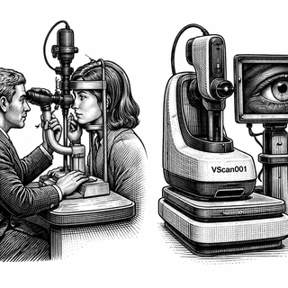
If you've ever sat in an ophthalmologist's office and pressed your chin into that little metal cradle while someone shines what feels like the surface of the sun directly into your eyeball, congratulations - you've experienced the slit lamp exam. It's the bread and butter of eye diagnostics, the sourdough starter of ophthalmology. And for roughly a century, it has required a trained human being to operate.
Lightfield Medical apparently looked at that arrangement and thought: "What if we just... didn't need the human part?"
Their clinical trial (NCT07495345) is now testing a device called the Vscan001 - an automated slit lamp camera that uses high-speed liquid lens technology to image the front of your eye. No trained photographer. No ophthalmologist hunched over the instrument squinting at your cornea. Just you, the machine, and whatever it finds in there.
What Exactly Is a Slit Lamp, and Why Should You Care?
For the uninitiated, the slit lamp is the workhorse of any eye clinic. It's essentially a high-powered microscope paired with a thin beam of light (the "slit") that lets a doctor examine the anterior segment of the eye - the cornea, iris, lens, and all the little structures up front that keep your vision from going sideways.
Think of it as slicing a very thin piece of sashimi off a block of tuna. The narrow beam of light creates an optical cross-section through the eye's structures, revealing details invisible to the naked eye. Cataracts, corneal ulcers, signs of glaucoma, infections - a skilled operator at a slit lamp can catch a remarkable number of problems before they become serious.
The catch? "Skilled operator" is doing a lot of heavy lifting in that sentence. High-quality slit lamp photography has traditionally been a specialized skill. There are more people on this planet who can make a decent croissant from scratch than can take publication-quality slit lamp images. The lamination of skill, equipment, and clinical knowledge required has created a real bottleneck in eye care delivery, particularly in underserved communities where ophthalmologists are about as common as Michelin-starred restaurants.
The Liquid Lens: Not What You'd Pour on a Salad
The secret ingredient in the Vscan001 is its high-speed liquid lens technology. If you're not familiar with liquid lenses, prepare to have your mind mildly rearranged.
A liquid lens uses a small volume of optical fluid whose shape can be changed electrically. Apply a voltage, the curvature of the liquid surface shifts, and the focal point changes - all without moving a single mechanical part. It's like adjustable-focus eyeglasses, except the adjustment happens in milliseconds rather than the time it takes you to find your reading glasses in the junk drawer.
This matters enormously for automated imaging. Traditional slit lamp photography requires the operator to manually adjust focus, illumination angle, and magnification in real-time based on what they're seeing. Automating that process with conventional optics would be like trying to automate a sushi chef's knife work using a series of fixed blades - technically possible, but slow and clumsy. Liquid lenses let the device rapidly sweep through focal planes, capturing sharp images across different depths of the eye faster than any human operator could adjust a dial.
Research into automated ophthalmic imaging has been accelerating over the past several years. Studies such as Balyen and Peto's 2019 review on AI applications in ophthalmology (DOI: 10.1007/s10792-019-01077-2) have highlighted how machine learning and automated imaging could transform diagnostic workflows. Meanwhile, work by Ting et al. on AI for detecting diabetic retinopathy and glaucoma (DOI: 10.1136/bjophthalmol-2018-313173) has shown that automated systems can match or exceed human performance in specific screening tasks - but only when they have quality images to work with. The Vscan001 is essentially trying to solve the upstream problem: getting those quality images in the first place, without requiring a specialist behind the camera.
The Feasibility Question: Can This Thing Actually Work in the Wild?
Here's where my inner skeptic orders a double espresso and settles in.
This is a feasibility study. That means Lightfield Medical isn't claiming the Vscan001 will replace your eye doctor. Not yet. They're asking a much more fundamental question: can this device successfully image a reasonable proportion of patients who walk into a real clinical setting?
That might sound like a low bar, but it's the right question to ask first. Eyes are not standardized widgets rolling off an assembly line. They come in wildly different configurations - deep-set eyes, narrow palpebral fissures, patients who can't hold still, patients who blink like they're sending Morse code. An automated system has to handle the full buffet of human ocular anatomy, not just the idealized test conditions of an engineering lab.
The trial enrolled adult patients at a single ophthalmic practice, with the primary endpoint being the proportion of patients who could be successfully imaged. No multi-site fireworks, no head-to-head against experienced photographers (that comes later, presumably). Just the fundamental question: does this recipe work with real ingredients, or only in the test kitchen?
Why This Matters More Than You Think
The global shortage of eye care professionals is not a theoretical problem. The World Health Organization estimates that over a billion people worldwide live with preventable vision impairment. Many of them lack access to even basic screening, let alone the specialized imaging that catches conditions early.
If the Vscan001 works - and that "if" is still earning its paycheck - it could slot into primary care offices, pharmacies, rural health clinics, and teleophthalmology networks where a trained slit lamp photographer is simply not available. A device that a medical assistant can set up, that patients can sit in front of, and that produces diagnostic-quality images without expert operation? That's not just an incremental improvement. That's moving the kitchen to where the diners are.
Yeung et al.'s work on smartphone-based anterior segment photography (DOI: 10.1016/j.jcjo.2020.01.001) showed promising results for low-cost imaging alternatives, but smartphone approaches generally sacrifice the controlled illumination and magnification that make slit lamp imaging so diagnostically powerful. The Vscan001 seems to be aiming for the best of both worlds - automation and accessibility without giving up image quality.
The Bottom Line
I've seen enough "revolutionary" medical devices to fill a landfill, and most of them deserved to end up there. But the Vscan001 is interesting precisely because it's not trying to boil the ocean. It's a feasibility study. It's asking the boring, essential question that too many device companies skip in their rush to slap "AI-powered" on a press release.
Can the machine take the picture? That's it. That's the whole trial.
And honestly? That's exactly the question that needs answering. Because if the automated camera can reliably image patients across the messy spectrum of real clinical encounters, everything downstream - AI diagnostics, teleophthalmology, screening programs in underserved communities - becomes dramatically more feasible.
I'll be watching this one like a chef watching a soufflé through the oven window. Cautiously optimistic, but fully prepared for collapse.
Trial Information: NCT07495345 | Table View
References:
- Balyen L, Peto T. Promising Artificial Intelligence-Machine Learning-Deep Learning Algorithms in Ophthalmology. Asia Pac J Ophthalmol. 2019;8(3):264-272. DOI: 10.1007/s10792-019-01077-2
- Ting DSW, et al. Artificial intelligence and deep learning in ophthalmology. Br J Ophthalmol. 2019;103(2):167-175. DOI: 10.1136/bjophthalmol-2018-313173
- Yeung WK, et al. Smartphone anterior segment photography. Can J Ophthalmol. 2020;55(3):e105-e108. DOI: 10.1016/j.jcjo.2020.01.001
Disclaimer: This blog post is for informational and educational purposes only. It does not constitute medical advice, diagnosis, or treatment recommendations. Clinical trials are ongoing research - outcomes are not guaranteed. Always consult a qualified healthcare professional for medical decisions. The views expressed are those of the author and do not necessarily reflect those of Biomedical Observer.