
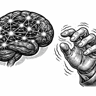
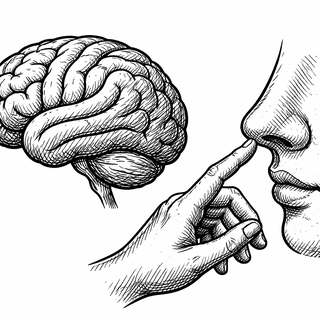
Try this: reach out and touch your nose. Easy, right? Now imagine that your hand shakes uncontrollably every time you try, or your arm decides to twist in a direction you didn't intend, or your finger just... stops working mid-reach. For millions of people with movement disorders, these seemingly simple actions become daily battles against a nervous system that won't cooperate.
Clinical trial NCT01019343 - Physiological Investigations of Movement Disorders - is the kind of study that doesn't get much press attention. It's not testing a glamorous new drug or a futuristic device. Instead, it's doing something more fundamental: mapping how movement disorders actually work at the level of brain circuits and electrical signals. And that groundwork might be more valuable than any individual treatment.
The NIH's Long Game
Run by the National Institute of Neurological Disorders and Stroke (NINDS), this study takes a different approach than your typical clinical trial. Rather than testing a specific intervention, it serves as a research platform - a way for investigators to probe the pathophysiology of movement disorders using non-invasive techniques.
The study design is flexible by intention. Researchers conduct pilot sub-studies that are exploratory, designed to generate hypotheses rather than prove them. When a small project leads to interesting results, a separate protocol with proper power analysis follows. It's the scientific equivalent of sending scouts ahead before committing the army.
What I find refreshing about this approach is its honesty about how science actually works. Most breakthroughs don't come from eureka moments - they come from careful observation, pattern recognition, and gradual accumulation of understanding. By creating a framework for systematically studying patients with movement disorders, NINDS is investing in the foundation that future treatments will be built upon.
The Cast of Characters: Movement Disorders 101
Movement disorders encompass a wide range of conditions, and this study doesn't discriminate. Patients with various diagnoses participate alongside healthy volunteers who serve as comparisons. Some of the major players include:
Parkinson's disease: The poster child of movement disorders, characterized by tremor, rigidity, and slowed movement (bradykinesia). It results from loss of dopamine-producing neurons in a brain region called the substantia nigra. About a million Americans live with Parkinson's, and that number is growing as the population ages.
Dystonia: A condition where muscles contract involuntarily, causing twisting, repetitive movements or abnormal postures. Dystonia can affect a single body part (like writer's cramp, which specifically targets hand muscles during writing) or be generalized. The pathophysiology involves abnormal signaling in the basal ganglia-thalamo-cortical circuits - which is a fancy way of saying the brain's motor control centers aren't talking to each other properly.
Essential tremor: The most common movement disorder, affecting up to 5% of people over 65. Despite the name suggesting it's somehow necessary (it's not), essential tremor causes rhythmic shaking, usually in the hands, that worsens during purposeful movement. The cerebellum and its connections play a key role.
Functional movement disorders: Perhaps the most fascinating and misunderstood category. These are movement abnormalities that aren't explained by damage to brain structure - the hardware looks fine, but the software isn't running right. They're not fake or imagined, but they arise from problems with how the brain processes movement rather than from neurodegeneration or injury.
The Tools of the Trade
What makes NCT01019343 particularly interesting is its toolkit. The study employs multiple non-invasive techniques to peer into brain function:
MRI (Magnetic Resonance Imaging): Beyond just taking pictures, modern MRI can reveal brain activity (functional MRI), map neural connections (diffusion tensor imaging tractography), measure regional brain volumes (voxel-based morphometry), and even detect levels of specific neurotransmitters (magnetic resonance spectroscopy). The brain is no longer a black box.
EEG (Electroencephalography): Those silly-looking caps covered with electrodes measure electrical activity across the scalp with millisecond precision. While MRI tells you where something happens, EEG tells you when. Combining the two gives researchers a surprisingly detailed picture of brain dynamics.
MEG (Magnetoencephalography): Similar to EEG but measuring magnetic fields rather than electrical potentials. MEG has better spatial resolution than EEG - think of it as the high-definition version. The downside? The equipment costs millions and requires magnetically shielded rooms.
TMS (Transcranial Magnetic Stimulation): Here TMS isn't being used as a treatment but as a probe. By delivering precisely timed magnetic pulses to specific brain regions, researchers can study connectivity and excitability. Fire a pulse at the motor cortex and measure how quickly and strongly the signal reaches the muscles - that tells you something about the pathway's integrity.
Peripheral nerve stimulation: Sometimes you need to work from the outside in. Stimulating peripheral nerves and measuring responses can reveal problems in the sensory feedback systems that help guide movement.
What They've Learned So Far
Decades of research using these techniques - including work from this study - have transformed our understanding of movement disorders:
Parkinson's disease: The bradykinesia characteristic of Parkinson's isn't just muscle weakness or slowness - it reflects abnormal patterns of activity in the motor cortex. Brain oscillations at specific frequencies (particularly beta waves around 20 Hz) become exaggerated, essentially putting the brakes on movement initiation.
Dystonia: Research has identified loss of inhibition and abnormal plasticity as key features. The brain of someone with dystonia is too "excitable" - circuits that should dampen inappropriate motor signals don't work properly. Additionally, the rules governing motor learning seem to be different, with maladaptive plasticity potentially contributing to the condition.
Essential tremor: The cerebellum emerges as the central player, with functional imaging showing abnormal cerebellar activity and altered connectivity in the cerebello-thalamo-cortical circuit. TMS studies have helped differentiate essential tremor from parkinsonian tremor based on which brain networks show abnormalities.
Functional movement disorders: Electrophysiological studies can actually help distinguish functional movement disorders from organic ones. Specific patterns - like the presence of a "Bereitschaftspotential" (readiness potential) before jerky movements - suggest involuntary movements that are generated through normal motor pathways, just not consciously controlled.
Why Basic Research Matters
I can already hear the impatient voice asking: "So what? When does this help actual patients?" Fair question, but let me make the case for why this kind of research matters:
Better diagnosis: Many movement disorders look similar on the surface. A tremor is a tremor, right? Wrong. The pattern, frequency, and circumstances of tremors differ between conditions, and understanding the underlying physiology helps clinicians tell them apart. Misdiagnosis in this field is surprisingly common - and it leads to wrong treatments.
Targeted treatments: If you know that a condition involves overactive beta oscillations, you can design interventions specifically targeting that abnormality. Deep brain stimulation settings can be optimized based on physiological understanding. New drugs can be developed to address specific circuit dysfunctions.
Measuring outcomes: How do you know if a treatment is working? Physiological measures provide objective markers beyond just asking patients "do you feel better?" This is particularly valuable in clinical trials where placebo effects can be substantial.
Identifying subtypes: Not everyone with the same diagnosis responds to the same treatment. Physiological profiling might eventually allow us to match patients with the interventions most likely to help them - personalized medicine for movement disorders.
The Scale of Ambition
NCT01019343 aims to enroll 1,200 healthy volunteers and 1,000 patients with movement disorders. That's a substantial commitment of resources, but it's necessary for the kind of systematic characterization the field needs. Individual small studies can be misleading - large databases of physiological data allow for more robust conclusions.
The exclusion criteria are practical: no metal implants that would interfere with MRI, ability to lie still for up to three hours (try that with a movement disorder - it's not easy), and no abnormalities on brain imaging unrelated to the diagnosis. Pregnancy also excludes participation, as it does for most imaging studies.
Participants undergo various combinations of the available techniques depending on the specific sub-study. Some might get an MRI session. Others might do TMS testing. The modular design allows researchers to mix and match based on their hypotheses.
The People Behind the Research
The study is led by Mark Hallett, M.D., Chief of the Human Motor Control Section at NINDS. Dr. Hallett has been a towering figure in movement disorder research for decades, with contributions spanning basic physiology to clinical applications. His work on cortical excitability in dystonia, the neurophysiology of psychogenic movement disorders, and the mechanisms of bradykinesia has influenced how a generation of neurologists thinks about these conditions.
Having researchers of this caliber dedicated to systematic physiological investigation is a luxury that patients might not appreciate but ultimately benefits everyone. The papers published from this research become reference standards that clinicians and researchers worldwide use to understand and treat movement disorders.
What Comes Next
Movement disorder research stands at an interesting crossroads. On one hand, we have more tools than ever to probe brain function non-invasively. On the other hand, translating that understanding into treatments remains challenging. The gap between knowing what goes wrong and knowing how to fix it is often larger than we'd like.
But that gap is shrinking. Physiological insights are increasingly guiding therapeutic development. Closed-loop deep brain stimulation - where the device adjusts its output based on real-time brain signals - represents the kind of precision medicine that basic research enables. New targets for neuromodulation emerge from careful circuit mapping.
For patients living with tremor, dystonia, or other movement disorders today, studies like NCT01019343 offer hope that science is steadily chipping away at the mystery of what's happening in their brains. It may not be glamorous work, but it's necessary work. And someday, when a new treatment emerges that finally helps, remember that it started with researchers patiently mapping brain signals one patient at a time.
References:
- ClinicalTrials.gov Identifier: NCT01019343
- National Institute of Neurological Disorders and Stroke (NINDS) - Physiological Investigations of Movement Disorders
- "Milestones in Clinical Neurophysiology" - PMC3105364
- "Frontiers in Human Neuroscience: Tapping the Potential of Multimodal Non-invasive Brain Stimulation to Elucidate the Pathophysiology of Movement Disorders"
Disclaimer: This blog post is for educational purposes only and does not constitute medical advice. If you are experiencing movement problems or have been diagnosed with a movement disorder, please consult with a qualified neurologist for appropriate evaluation and management. Research studies involve eligibility criteria and should only be participated in after careful consideration and informed consent. Images and graphics are for illustrative purposes only and do not depict actual medical devices, procedures, mechanisms, or research findings from the referenced studies.